25.1: The Human Eye
- Page ID
- 16186
\( \newcommand{\vecs}[1]{\overset { \scriptstyle \rightharpoonup} {\mathbf{#1}} } \)
\( \newcommand{\vecd}[1]{\overset{-\!-\!\rightharpoonup}{\vphantom{a}\smash {#1}}} \)
\( \newcommand{\dsum}{\displaystyle\sum\limits} \)
\( \newcommand{\dint}{\displaystyle\int\limits} \)
\( \newcommand{\dlim}{\displaystyle\lim\limits} \)
\( \newcommand{\id}{\mathrm{id}}\) \( \newcommand{\Span}{\mathrm{span}}\)
( \newcommand{\kernel}{\mathrm{null}\,}\) \( \newcommand{\range}{\mathrm{range}\,}\)
\( \newcommand{\RealPart}{\mathrm{Re}}\) \( \newcommand{\ImaginaryPart}{\mathrm{Im}}\)
\( \newcommand{\Argument}{\mathrm{Arg}}\) \( \newcommand{\norm}[1]{\| #1 \|}\)
\( \newcommand{\inner}[2]{\langle #1, #2 \rangle}\)
\( \newcommand{\Span}{\mathrm{span}}\)
\( \newcommand{\id}{\mathrm{id}}\)
\( \newcommand{\Span}{\mathrm{span}}\)
\( \newcommand{\kernel}{\mathrm{null}\,}\)
\( \newcommand{\range}{\mathrm{range}\,}\)
\( \newcommand{\RealPart}{\mathrm{Re}}\)
\( \newcommand{\ImaginaryPart}{\mathrm{Im}}\)
\( \newcommand{\Argument}{\mathrm{Arg}}\)
\( \newcommand{\norm}[1]{\| #1 \|}\)
\( \newcommand{\inner}[2]{\langle #1, #2 \rangle}\)
\( \newcommand{\Span}{\mathrm{span}}\) \( \newcommand{\AA}{\unicode[.8,0]{x212B}}\)
\( \newcommand{\vectorA}[1]{\vec{#1}} % arrow\)
\( \newcommand{\vectorAt}[1]{\vec{\text{#1}}} % arrow\)
\( \newcommand{\vectorB}[1]{\overset { \scriptstyle \rightharpoonup} {\mathbf{#1}} } \)
\( \newcommand{\vectorC}[1]{\textbf{#1}} \)
\( \newcommand{\vectorD}[1]{\overrightarrow{#1}} \)
\( \newcommand{\vectorDt}[1]{\overrightarrow{\text{#1}}} \)
\( \newcommand{\vectE}[1]{\overset{-\!-\!\rightharpoonup}{\vphantom{a}\smash{\mathbf {#1}}}} \)
\( \newcommand{\vecs}[1]{\overset { \scriptstyle \rightharpoonup} {\mathbf{#1}} } \)
\(\newcommand{\longvect}{\overrightarrow}\)
\( \newcommand{\vecd}[1]{\overset{-\!-\!\rightharpoonup}{\vphantom{a}\smash {#1}}} \)
\(\newcommand{\avec}{\mathbf a}\) \(\newcommand{\bvec}{\mathbf b}\) \(\newcommand{\cvec}{\mathbf c}\) \(\newcommand{\dvec}{\mathbf d}\) \(\newcommand{\dtil}{\widetilde{\mathbf d}}\) \(\newcommand{\evec}{\mathbf e}\) \(\newcommand{\fvec}{\mathbf f}\) \(\newcommand{\nvec}{\mathbf n}\) \(\newcommand{\pvec}{\mathbf p}\) \(\newcommand{\qvec}{\mathbf q}\) \(\newcommand{\svec}{\mathbf s}\) \(\newcommand{\tvec}{\mathbf t}\) \(\newcommand{\uvec}{\mathbf u}\) \(\newcommand{\vvec}{\mathbf v}\) \(\newcommand{\wvec}{\mathbf w}\) \(\newcommand{\xvec}{\mathbf x}\) \(\newcommand{\yvec}{\mathbf y}\) \(\newcommand{\zvec}{\mathbf z}\) \(\newcommand{\rvec}{\mathbf r}\) \(\newcommand{\mvec}{\mathbf m}\) \(\newcommand{\zerovec}{\mathbf 0}\) \(\newcommand{\onevec}{\mathbf 1}\) \(\newcommand{\real}{\mathbb R}\) \(\newcommand{\twovec}[2]{\left[\begin{array}{r}#1 \\ #2 \end{array}\right]}\) \(\newcommand{\ctwovec}[2]{\left[\begin{array}{c}#1 \\ #2 \end{array}\right]}\) \(\newcommand{\threevec}[3]{\left[\begin{array}{r}#1 \\ #2 \\ #3 \end{array}\right]}\) \(\newcommand{\cthreevec}[3]{\left[\begin{array}{c}#1 \\ #2 \\ #3 \end{array}\right]}\) \(\newcommand{\fourvec}[4]{\left[\begin{array}{r}#1 \\ #2 \\ #3 \\ #4 \end{array}\right]}\) \(\newcommand{\cfourvec}[4]{\left[\begin{array}{c}#1 \\ #2 \\ #3 \\ #4 \end{array}\right]}\) \(\newcommand{\fivevec}[5]{\left[\begin{array}{r}#1 \\ #2 \\ #3 \\ #4 \\ #5 \\ \end{array}\right]}\) \(\newcommand{\cfivevec}[5]{\left[\begin{array}{c}#1 \\ #2 \\ #3 \\ #4 \\ #5 \\ \end{array}\right]}\) \(\newcommand{\mattwo}[4]{\left[\begin{array}{rr}#1 \amp #2 \\ #3 \amp #4 \\ \end{array}\right]}\) \(\newcommand{\laspan}[1]{\text{Span}\{#1\}}\) \(\newcommand{\bcal}{\cal B}\) \(\newcommand{\ccal}{\cal C}\) \(\newcommand{\scal}{\cal S}\) \(\newcommand{\wcal}{\cal W}\) \(\newcommand{\ecal}{\cal E}\) \(\newcommand{\coords}[2]{\left\{#1\right\}_{#2}}\) \(\newcommand{\gray}[1]{\color{gray}{#1}}\) \(\newcommand{\lgray}[1]{\color{lightgray}{#1}}\) \(\newcommand{\rank}{\operatorname{rank}}\) \(\newcommand{\row}{\text{Row}}\) \(\newcommand{\col}{\text{Col}}\) \(\renewcommand{\row}{\text{Row}}\) \(\newcommand{\nul}{\text{Nul}}\) \(\newcommand{\var}{\text{Var}}\) \(\newcommand{\corr}{\text{corr}}\) \(\newcommand{\len}[1]{\left|#1\right|}\) \(\newcommand{\bbar}{\overline{\bvec}}\) \(\newcommand{\bhat}{\widehat{\bvec}}\) \(\newcommand{\bperp}{\bvec^\perp}\) \(\newcommand{\xhat}{\widehat{\xvec}}\) \(\newcommand{\vhat}{\widehat{\vvec}}\) \(\newcommand{\uhat}{\widehat{\uvec}}\) \(\newcommand{\what}{\widehat{\wvec}}\) \(\newcommand{\Sighat}{\widehat{\Sigma}}\) \(\newcommand{\lt}{<}\) \(\newcommand{\gt}{>}\) \(\newcommand{\amp}{&}\) \(\definecolor{fillinmathshade}{gray}{0.9}\)learning objectives
- Identify parts of human eye and their functions
The human eye is the gateway to one of our five senses. The human eye is an organ that reacts with light. It allows light perception, color vision and depth perception. A normal human eye can see about 10 million different colors! There are many parts of a human eye, and that is what we are going to cover in this atom.
Properties
Contrary to what you might think, the human eye is not a perfect sphere, but is made up of two differently shaped pieces, the cornea and the sclera. These two parts are connected by a ring called the limbus. The part of the eye that is seen is the iris, which is the colorful part of the eye. In the middle of the iris is the pupil, the black dot that changes size. The cornea covers these elements, but is transparent. The fundus is on the opposite of the pupil, but inside the eye and can not be seen without special instruments. The optic nerve is what conveys the signals of the eye to the brain. is a diagram of the eye. The human eye is made up of three coats:
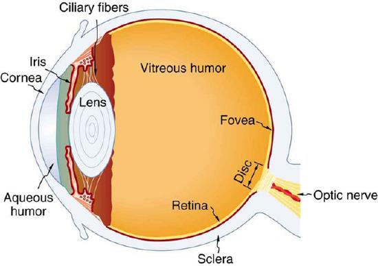
Diagram of the Human Eye: The cornea and lens of an eye act together to form a real image on the light-sensing retina, which has its densest concentration of receptors in the fovea and a blind spot over the optic nerve. The power of the lens of an eye is adjustable to provide an image on the retina for varying object distances. Layers of tissues with varying indices of refraction in the lens are shown here. However, they have been omitted from other pictures for clarity.
- Outermost Layer – composed of the cornea and the sclera.
- Middle Layer – composed of the choroid, ciliary body and iris.
- Innermost Layer – the retina, which can be seen with an instrument called the ophthalmoscope.
Once you are inside these three layers, there is the aqueous humor (clear fluid that is contained in the anterior chamber and posterior chamber), vitreous body (clear jelly that is much bigger than the aqueous humor), and the flexible lens. All of these are connected by the pupil.
Dynamics
Whenever the eye moves, even just a little, it automatically readjusts the exposure by adjusting the iris, which regulates the size of the pupil. This is what helps the eye adjust to dark places or really bright lights. The lens of the eye is similar to one in glasses or cameras. The human eye is had an aperture, just like a camera. The pupil serves this function, and the iris is the aperture stop. The different parts of the eye has different refractive indexes, and this is what bends the rays to form an image. The cornea provides two-thirds of the power to the eye. The lens provides the remaining power. The image passes through several layers of the eye, but happens in a way very similar to that of a convex lens. When the image finally reaches the retena, it is inverted, but the brain will correct this. shows what happens.
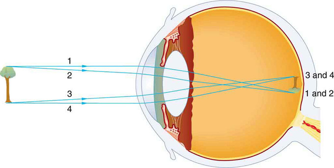
Vision Diagram: An image is formed on the retina with light rays converging most at the cornea and upon entering and exiting the lens. Rays from the top and bottom of the object are traced and produce an inverted real image on the retina. The distance to the object is drawn smaller than scale.
Eye Movement
Each eye has six muscles; lateral rectus, medial rectus, inferior rectus, superior rectus, inferior oblique, and superior oblique. All of these muscles provide differnt tensions and torques to control the movement of the eye. These are a few examples of types of eye movement:
- Rapid Eye Movement – Often referred to as REM, this happens in the sleep stage when most vivid dreams occur.
- Saccade – These are quick, simultaneous movements of both eyes, and is controlled by the frontal lobe of the brain.
- Vestibulo-ocular Reflex – This is the eye movement which is opposite to the movement of the head and keeps the object you are looking at in the center of vision.
- Pursuit Movement – This is the tracking movement when you are following a moving object. It is less accurate than the vestibulo-ocular reflex.
Color Vision
Using the cone cells in the retina, we perceive images in color; each type of cone specifically sees in regions of red, green, or blue.
learning objectives
- Explain how the human eye perceives colors
With human eyesight, cone cells are responsible for color vision. From there, it is important to understand how color is perceived. Using the cone cells in the retina, we perceive images in color. Each type of cone specifically sees in regions of red, green, or blue, (RGB), in the color spectrum of red, orange, yellow, green, blue, indigo, violet.
The colors in between these absolutes are seen as different linear combinations of RGB. This is why TVs and computer screens are made up of thousands of little red, green, or blue lights, and why colors in electronic form are represented by different values of RGB. These values are usually given in the value of their frequency in log form.
YUV Color Space
The human eye is more sensitive to intensity changes than color changes, which is why it is acceptable to use black and white photography in place of color and why people can still distinguish everything in the photo without colors. The intensity, or luminance Y, can be found from the following equation:
\[\left. \begin{array} { l } { \mathrm { Y } = 0.3 \mathrm { R } + 0.6 \mathrm { G } + 0.1 \mathrm { B } } \\ { \mathrm { Y } = 0.3 \mathrm { R } + 0.6 \mathrm { G } + 0.1 \mathrm { B } } \end{array} \right.\]
The prior equation deals with the luminance, but the chrominance (dealing with colors) can be found from the following equations:
\[\left. \begin{array} { l } { \mathrm { U } = 0.5 ( \mathrm { BY } ) } \\ { \mathrm { U } = 0.5 ( \mathrm { B } - \mathrm { Y } ) } \\ { \mathrm { V } = 0.625 ( \mathrm { R } - \mathrm { Y } ) } \end{array} \right.\]
\[\mathrm { V } = 0.625 ( \mathrm { R } \mathrm { Y } )\]
You can go from RGB to YUV color spaces with the following matrix operation:
\[\left( \begin{array} { c } { \mathrm { Y } } \\ { \mathrm { U } } \\ { \mathrm { V } } \end{array} \right) = \mathrm { C } * \left( \begin{array} { l } { \mathrm { R } } \\ { \mathrm { G } } \\ { \mathrm { B } } \end{array} \right)\]
Where C is equal to:
\[\left( \begin{array} { c c c } { 0.3 } & { 0.6 } & { 0.1 } \\ { - 0.15 } & { - 0.3 } & { 0.45 } \\ { 0.4375 } & { - 0.3750 } & { - 0.0625 } \end{array} \right)\]
Visual Sensitivity
In, we can see that
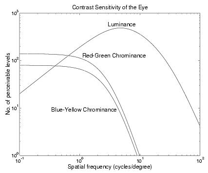
Visual Sensitivity: This graph shows the sensitivity of the eye to luminance (Y) and chrominance (U, V) components of images. The horizontal scale is spatial frequency, and represents the frequency of an alternating pattern of parallel stripes with sinusoidally varying intensity. The vertical scale is the contrast sensitivity of human vision, which is the ratio of the maximum visible range of intensities to the minimum discernible peak-to-peak intensity variation at the specified frequency.
- the maximum sensitivity to Y occurs for spatial frequencies around 5 cycles / degree, which corresponds to striped patterns with a half-period (stripe width) of 1.8 mm at a distance of 1 m (~arm’s length).
- The eye has very little response above 100 cycles / degree, which corresponds to a stripe width of 0.1 mm at 1 m. On a standard PC display of width 250 mm, this would require 2500 pels per line! Hence the current SVGA standard of 1024×768 pels still falls somewhat short of the ideal and is limited by CRT spot size. Modern laptop displays have a pel size of about 0.3 mm, but are pleasing to view because the pel edges are so sharp (and there is no flicker).
- The sensitivity to luminance drops off at low spatial frequencies, showing that we are not very good at estimating absolute luminance levels as long as they do not change with time – the luminance sensitivity to temporal fluctuations (flicker) does not fall off at low spatial frequencies.
- The maximum chrominance sensitivity is much lower than the maximum luminance sensitivity with blue-yellow (U) sensitivity being about half of red-green (V) sensitivity and about 16 of the maximum luminance sensitivity.
- The chrominance sensitivities fall off above 1 cycle / degree, requiring a much lower spatial bandwidth than luminance.
We can now see why it is better to convert to the YUV domain before attempting image compression. The U and V components may be sampled at a lower rate than Y (due to narrower bandwidth) and may be quantified more coarsely (due to lower contrast sensitivity).
Resolution of the Human Eye
The human eye is a sense organ that allows vision and is capable to distinguish about 10 million colors.
learning objectives
- Describe field of view and color sensitivity of the human eye
The human eye is an organ that reacts to light in many circumstances. As a conscious sense organ the human eye allows vision; rod and cone cells in the retina allow conscious light perception and vision, including color differentiation and the perception of depth. The human eye can distinguish about 10 million colors. A model of the human eye can be seen in.
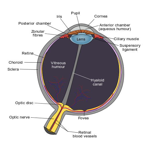
Schematic Diagram of the Human Eye: Structure of the eye and closeup of the retina.
The retina of human eye has a static contrast ratio of around 100:1 (about 6.5 f-stops). As soon as the eye moves, it re-adjusts its exposure, both chemically and geometrically, by adjusting the iris (which regulates the size of the pupil ). Initial dark adaptation takes place in approximately four seconds of profound, uninterrupted darkness; full adaptation, through adjustments in retinal chemistry, is mostly complete in thirty minutes. Hence, a dynamic contrast ratio of about 1,000,000:1 (about 20 f-stops) is possible. The process is nonlinear and multifaceted, so an interruption by light starts the adaptation process over again. Full adaptation is dependent on good blood flow (thus dark adaptation may be hampered by poor circulation, and vasoconstrictors like tobacco).
The eye includes a lens not dissimilar to lenses found in optical instruments (such as cameras). The same principles can be applied. The pupil of the human eye is its aperture. The iris is the diaphragm that serves as the aperture stop. Refraction in the cornea causes the effective aperture (the entrance pupil) to differ slightly from the physical pupil diameter. The entrance pupil is typically about 4 mm in diameter, although it can range from 2 mm (f/8.3) in a brightly lit place to 8 mm (f/2.1) in the dark. The latter value decreases slowly with age; older people’s eyes sometimes dilate to not more than 5-6mm.
The approximate field of view of an individual human eye is 95° away from the nose, 75° downward, 60° toward the nose, and 60° upward, allowing humans to have an almost 180-degree forward-facing horizontal field of view. With eyeball rotation of about 90° (head rotation excluded, peripheral vision included), horizontal field of view is as high as 170°. About 12–15° temporal and 1.5° below the horizontal is the optic nerve or blind spot which is roughly 7.5° high and 5.5° wide.
Nearsightedness, Farsidedness, and Vision Correction
In order for the human eye to see clearly, the image needs to be formed directly on the retina; if it is not, the image is blurry.
learning objectives
- Identify factors responsible for nearsightedness and farsightedness vision defects
The human eye is the gateway to one of our five senses. The human eye is an organ that reacts with light. It allows light perception, color vision, and depth perception, but not all eyes are perfect. A normal human eye can see about 10 million different colors!
Properties
Contrary to what you might think, the human eye is not a perfect sphere, but is made up of two differently shaped pieces, the cornea and the sclera. These two parts are connected by a ring called the limbus. The part of the eye that is seen is the iris, which is the colorful part of the eye. In the middle of the iris is the pupil, which is the black dot that changes size. The cornea covers these elements, but it is transparent. The fundus is on the opposite of the pupil, but inside the eye and cannot be seen without special instruments. The optic nerve is what conveys the signals of the eye to the brain. shows a diagram of the eye.
Vision
The different parts of the eye have different refractive indexes, and this is what bends the rays to form an image. The cornea provides two-thirds of the power to the eye. The lens provides the remaining power. The image passes through several layers of the eye, but this happens in a way very similar to that of a convex lens. When the image finally reaches the retena, it is inverted, but the brain will correct this. For the vision to be clear, the image has to be formed directly on the retina. The focus needs to be changed, much like a camera, depending on the distance and size of the object. The eye’s lens is flexible, and changes shape. This changes the focal length. The eye’s ciliary muscles control the shape of the lens. When you focus on something, you squeeze or relax these muscles.
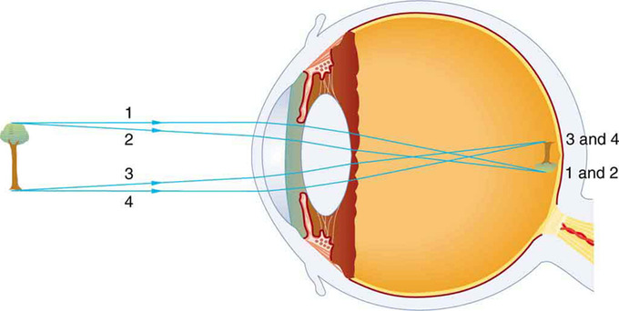
Vision Diagram: An image is formed on the retina with light rays converging most at the cornea and upon entering and exiting the lens. Rays from the top and bottom of the object are traced and produce an inverted real image on the retina. The distance to the object is drawn smaller than scale.
Near Sighted Vision
Nearsightedness, or myopia is a vision defect that occurs when the focus of the image is in front of the retina. This is shown in. Close objects are seen fine, but distant objects are blurry. This can be corrected by placing diverging lenses in front of the eye. This will cause the light rays to spread out before they enter the eye.

Near Sighted Vision: This occurs when the image is formed before the retina
Far Sighted Vision
Farsightedness, or hyperopia, is a vision defect that occurs when the focus of the image is behind the retina. This is shown in. Distant objects are seen fine, but closer objects are blurry. This can be corrected by placing converging lenses in front of the eye. This will cause the light rays to slightly converge together before they enter the eye.

Far Sighted Vision: This occurs when the image is formed behind the retina
Key Points
- The eye is made up of a number of parts, including the iris, pupil, cornea, and retina.
- The eye has six muscles which control the eye movement, all providing different tension and torque.
- The eye works a lot like a camera, the pupil provides the f-stop, the iris the aperture stop, the cornea resembles a lens. The way that the image is formed is much like the way a convex lens forms an image.
- The cones in the retina are responsible for seeing colors. There are three types of cones, each type can only pick up one color: red, green, or blue. This is why TVs and computer screens are made up of thousands of little red, green, or blue lights.
- The human eye is more sensitive to intensity changes than color changes, which is why it is acceptable to use black and white photography in place of color and why people can still distinguish everything in the photo without colors.
- Colors are usually written in different values of red, green, or blue. Each value is the log form of that frequency.
- The retina of human eye has a static contrast ratio of around 100:1 and a dynamic contrast ratio of about 1,000,000:1.
- The eye includes a lens not dissimilar to lenses found in optical instruments, such as cameras.
- The approximate field of view of an individual human eye is 95° away from the nose, 75° downward, 60° toward the nose, and 60° upward, allowing humans to have an almost 180-degree forward-facing horizontal field of view.
- The focal point of the image will change depending on how the lens is shaped. Your lens changes depending on the distance of the object, by the relaxation or contraction of the muscles, and this controls the focal length.
- Near sightedness occurs when the image is formed before the retina.
- Farsightedness occurs when the image is formed behind the retina.
Key Terms
- pupil: The hole in the middle of the iris of the eye, through which light passes to be focused on the retina.
- aperture: The diameter of the aperture that restricts the width of the light path through the whole system. For a telescope, this is the diameter of the objective lens (e.g., a telescope may have a 100 cm aperture).
- luminance: The intensity of an object, independent from its color.
- static contrast ratio: Luminosity ratio of the brightest and darkest color the system is capable of processing simultaneously at any instant of time.
- dynamic contrast ratio: Luminosity ratio of the brightest and darkest color the system is capable of processing over time (while the picture is moving).
- field of view: The angular extent of what can be seen, either with the eye or with an optical instrument or camera.
- myopia: A disorder of the vision where distant objects appear blurred because the eye focuses their images in front of the retina instead of on it.
- hyperopia: A disorder of the vision where the eye focusses images behind the retina instead of on it, so that distant objects can be seen better than near objects.
LICENSES AND ATTRIBUTIONS
CC LICENSED CONTENT, SHARED PREVIOUSLY
- Curation and Revision. Provided by: Boundless.com. License: CC BY-SA: Attribution-ShareAlike
CC LICENSED CONTENT, SPECIFIC ATTRIBUTION
- Human eye. Provided by: Wikipedia. Located at: en.Wikipedia.org/wiki/Human_eye. License: CC BY-SA: Attribution-ShareAlike
- OpenStax College, Physics of the Eye. September 17, 2013. Provided by: OpenStax CNX. Located at: http://cnx.org/content/m42482/latest/. License: CC BY: Attribution
- aperture. Provided by: Wiktionary. Located at: en.wiktionary.org/wiki/aperture. License: CC BY-SA: Attribution-ShareAlike
- pupil. Provided by: Wiktionary. Located at: en.wiktionary.org/wiki/pupil. License: CC BY-SA: Attribution-ShareAlike
- OpenStax College, Physics of the Eye. December 25, 2012. Provided by: OpenStax CNX. Located at: http://cnx.org/content/m42482/latest/. License: CC BY: Attribution
- OpenStax College, Physics of the Eye. December 25, 2012. Provided by: OpenStax CNX. Located at: http://cnx.org/content/m42482/latest/. License: CC BY: Attribution
- Optics. Provided by: Wikipedia. Located at: en.Wikipedia.org/wiki/Optics. License: CC BY-SA: Attribution-ShareAlike
- Nick Kingsbury, Human Vision. September 18, 2013. Provided by: OpenStax CNX. Located at: http://cnx.org/content/m11084/latest/. License: CC BY: Attribution
- luminance. Provided by: Wikipedia. Located at: en.Wikipedia.org/wiki/luminance. License: CC BY-SA: Attribution-ShareAlike
- OpenStax College, Physics of the Eye. December 25, 2012. Provided by: OpenStax CNX. Located at: http://cnx.org/content/m42482/latest/. License: CC BY: Attribution
- OpenStax College, Physics of the Eye. December 25, 2012. Provided by: OpenStax CNX. Located at: http://cnx.org/content/m42482/latest/. License: CC BY: Attribution
- Nick Kingsbury, Human Vision. December 25, 2012. Provided by: OpenStax CNX. Located at: http://cnx.org/content/m11084/latest/. License: CC BY: Attribution
- field of view. Provided by: Wiktionary. Located at: en.wiktionary.org/wiki/field_of_view. License: CC BY-SA: Attribution-ShareAlike
- Optics. Provided by: Wikipedia. Located at: en.Wikipedia.org/wiki/Optics. License: CC BY-SA: Attribution-ShareAlike
- Human eye. Provided by: Wikipedia. Located at: en.Wikipedia.org/wiki/Human_e...3Dynamic_range. License: CC BY-SA: Attribution-ShareAlike
- static contrast ratio. Provided by: Wikipedia. Located at: en.Wikipedia.org/wiki/static%...ntrast%20ratio. License: CC BY-SA: Attribution-ShareAlike
- dynamic contrast ratio. Provided by: Wikipedia. Located at: en.Wikipedia.org/wiki/dynamic...ntrast%20ratio. License: CC BY-SA: Attribution-ShareAlike
- OpenStax College, Physics of the Eye. December 25, 2012. Provided by: OpenStax CNX. Located at: http://cnx.org/content/m42482/latest/. License: CC BY: Attribution
- OpenStax College, Physics of the Eye. December 25, 2012. Provided by: OpenStax CNX. Located at: http://cnx.org/content/m42482/latest/. License: CC BY: Attribution
- Nick Kingsbury, Human Vision. December 25, 2012. Provided by: OpenStax CNX. Located at: http://cnx.org/content/m11084/latest/. License: CC BY: Attribution
- File:Schematic diagram of the human eye en.svg - Wikibooks, open books for an open world. Provided by: Wikibooks. Located at: en.wikibooks.org/w/index.php?...20080202013345. License: CC BY-SA: Attribution-ShareAlike
- OpenStax College, Physics of the Eye. September 17, 2013. Provided by: OpenStax CNX. Located at: http://cnx.org/content/m42482/latest/. License: CC BY: Attribution
- Free High School Science Texts Project, Geometrical Optics: The Human Eye (Grade 11). September 17, 2013. Provided by: OpenStax CNX. Located at: http://cnx.org/content/m39031/latest/. License: CC BY: Attribution
- Human eye. Provided by: Wikipedia. Located at: en.Wikipedia.org/wiki/Human_eye. License: CC BY-SA: Attribution-ShareAlike
- hyperopia. Provided by: Wiktionary. Located at: en.wiktionary.org/wiki/hyperopia. License: CC BY-SA: Attribution-ShareAlike
- myopia. Provided by: Wiktionary. Located at: en.wiktionary.org/wiki/myopia. License: CC BY-SA: Attribution-ShareAlike
- OpenStax College, Physics of the Eye. December 25, 2012. Provided by: OpenStax CNX. Located at: http://cnx.org/content/m42482/latest/. License: CC BY: Attribution
- OpenStax College, Physics of the Eye. December 25, 2012. Provided by: OpenStax CNX. Located at: http://cnx.org/content/m42482/latest/. License: CC BY: Attribution
- Nick Kingsbury, Human Vision. December 25, 2012. Provided by: OpenStax CNX. Located at: http://cnx.org/content/m11084/latest/. License: CC BY: Attribution
- File:Schematic diagram of the human eye en.svg - Wikibooks, open books for an open world. Provided by: Wikibooks. Located at: en.wikibooks.org/w/index.php?...20080202013345. License: CC BY-SA: Attribution-ShareAlike
- Free High School Science Texts Project, Geometrical Optics: The Human Eye (Grade 11). December 25, 2012. Provided by: OpenStax CNX. Located at: http://cnx.org/content/m39031/latest/. License: CC BY: Attribution
- OpenStax College, Physics of the Eye. December 25, 2012. Provided by: OpenStax CNX. Located at: http://cnx.org/content/m42482/latest/. License: CC BY: Attribution
- Free High School Science Texts Project, Geometrical Optics: The Human Eye (Grade 11). December 25, 2012. Provided by: OpenStax CNX. Located at: http://cnx.org/content/m39031/latest/. License: CC BY: Attribution

